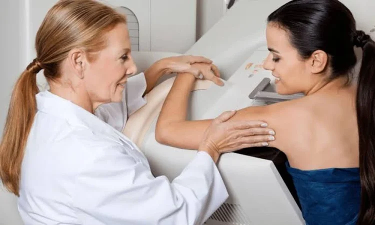
Finding a breast lump ranks among the most anxiety-inducing health discoveries. While most breast lumps prove benign, determining whether tissue changes warrant concern requires more than a physical exam. The path from detection to diagnosis involves multiple imaging technologies, each suited to specific circumstances and tissue types.
Mammography remains the foundation of breast screening, but when it reveals suspicious areas or when you can feel a lump that imaging cannot clearly characterize, additional diagnostic steps become necessary. Understanding when standard imaging falls short and what happens next helps reduce the uncertainty that accompanies abnormal findings.
How Breast Tissue Density Affects Detection
Breast tissue density varies considerably among women and changes throughout life. Dense breast tissue appears white on mammograms, as do masses, which creates a detection challenge similar to spotting a snowball in a snowstorm. Roughly 40 percent of women have heterogeneously dense or extremely dense breasts, meaning fibroglandular tissue occupies a larger proportion of the breast volume than fatty tissue.
Dense tissue does not indicate disease, but it does reduce mammography’s sensitivity. A 2016 study found that adding ultrasound screening in women with dense breasts detected an additional 3.2 cancers per 1,000 women screened, though it also increased false positives. The decision to use supplemental screening depends on breast density combined with other risk factors including family history, prior biopsies, and genetic predisposition.
Younger women typically have denser breast tissue, which gradually becomes more fatty with age and after menopause. Hormone replacement therapy can reverse this trend, maintaining or increasing density. This physiological reality means that detection strategies must adapt to individual tissue composition rather than relying on age-based protocols alone.
The Role of Ultrasound in Breast Evaluation
Ultrasound serves as the primary supplemental tool for evaluating palpable lumps and characterizing abnormalities seen on mammography. Unlike X-rays, ultrasound uses sound waves that penetrate dense tissue effectively, distinguishing fluid-filled cysts from solid masses. This distinction matters because simple cysts rarely require further action, while solid masses need additional assessment.
The technology excels at evaluating specific areas of concern but works less well as a standalone screening tool for the entire breast. Radiologists use standardized descriptors to classify ultrasound findings, noting features like shape, margin definition, orientation, and internal echo patterns. A mass described as having irregular margins, being taller than wide, or showing posterior acoustic shadowing raises suspicion and typically prompts biopsy recommendation.
Doppler ultrasound adds another dimension by assessing blood flow within masses. Malignant tumors often develop their own blood supply with characteristic flow patterns, though this finding alone cannot definitively separate benign from malignant tissue. The interpretation requires expertise, as overlapping features between benign and malignant lesions occur frequently.
When Tissue Sampling Becomes Necessary
Imaging provides morphological information but cannot make a definitive diagnosis. When findings suggest potential malignancy, obtaining tissue for microscopic examination becomes the critical next step. The traditional approach involved surgical excision, but this method has largely given way to less invasive techniques that provide diagnostic accuracy without requiring an operating room.
Core needle biopsy has become the standard for tissue diagnosis in breast lesions. Unlike fine needle aspiration which extracts individual cells, core biopsy removes intact tissue cylinders that preserve architectural patterns pathologists need to distinguish between different conditions. The procedure typically retrieves four to eight samples from different areas of the suspicious region, reducing sampling error.
For masses visible on ultrasound, an ultrasound guided biopsy offers real-time visualization throughout the procedure. The radiologist watches the needle enter the target lesion on the ultrasound screen, ensuring accurate sampling of the intended area. This approach works well for palpable lumps and sonographically visible abnormalities, typically performed with local anesthesia in an outpatient setting. Cleveland Clinic Abu Dhabi provides this diagnostic service as part of comprehensive breast care, allowing patients in the UAE to access the same protocols used in complex medical centers internationally.
What Biopsy Results Actually Mean
Pathology reports categorize breast biopsy findings into benign, high-risk, or malignant. Benign diagnoses like fibroadenoma or fibrocystic change require no further treatment, though imaging follow-up may be recommended to confirm stability. High-risk lesions including atypical ductal hyperplasia or lobular carcinoma in situ occupy a grey zone, associated with increased cancer risk but not representing cancer themselves. These findings often lead to surgical excision to ensure no adjacent malignancy was missed.
Malignant diagnoses trigger staging work-up and treatment planning. The pathology report specifies cancer type, grade, hormone receptor status, and HER2 status. These molecular markers guide therapy selection more precisely than cancer stage alone. A cancer that is estrogen receptor positive responds to different treatments than triple-negative disease, even when tumors are the same size and stage.
Occasionally, biopsy results return as non-diagnostic, meaning the sample contained insufficient tissue or missed the target lesion entirely. This outcome necessitates repeat biopsy, sometimes using a different guidance method. Persistence is essential because discordant results, where imaging suggests malignancy but pathology shows benign tissue, require resolution rather than assumption that the benign result is correct.
Managing the Emotional Weight of Investigation
The interval between discovering a lump and receiving biopsy results spans days to weeks, a period marked by uncertainty and anxiety. Research has shown that the psychological distress during this diagnostic phase can significantly impact wellbeing, sometimes persisting even after benign results provide reassurance.
Having a healthcare team that communicates clearly about what each test can and cannot determine reduces some of this anxiety. Understanding that multiple tests serve complementary rather than redundant purposes helps frame the diagnostic process as thorough rather than inefficient. Each imaging modality and biopsy technique answers specific questions that together build a complete picture.
For those managing diagnostic uncertainty, maintaining routine health behaviors helps restore a sense of control. Just as establishing consistent care relationships for family health needs provides continuity and reduces friction in healthcare access, developing a relationship with a breast imaging center that has your prior images and understands your baseline tissue patterns streamlines future evaluations.
Moving Forward After Diagnosis
A benign biopsy result does not necessarily mean returning to routine annual screening. Depending on what was found, imaging surveillance at six-month intervals may be recommended to document stability before reverting to annual schedules. High-risk lesions may warrant discussion with a breast surgeon about whether surgical excision or enhanced screening provides better risk management for your specific situation.
When biopsy confirms cancer, the focus shifts immediately to staging and treatment planning. Most women diagnosed with early-stage breast cancer today receive breast-conserving surgery rather than mastectomy, followed by radiation. Systemic therapy decisions depend on tumor biology more than size, with hormone-positive cancers treated differently than HER2-positive or triple-negative disease.
The diagnostic journey from finding a lump to definitive diagnosis requires patience with medical processes designed to avoid both missed cancers and unnecessary surgery. While technology continues advancing, with molecular imaging and artificial intelligence interpretation on the horizon, current diagnostic pathways balance sensitivity, specificity, and invasiveness effectively for most clinical scenarios. Understanding what each step accomplishes helps transform an anxious waiting period into an informed participation in your own care.